And It's About Time There Was Some Support For Cushing's!
From UCLA
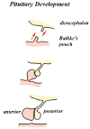
Rathke's pouch is a normal component of development which eventually forms the anterior lobe, pars intermedia and pars tuberalis of the pituitary gland. This pouch normally closes early in fetal development, but a remnant often persists as a cleft that lies within the pituitary gland. Occasionally, this remnant gives rise to a large cyst, the Rathke's cleft cyst (RCC), that causes symptoms.
Symptomatic RCCs are relatively uncommon lesions, accounting for less than 1% of all primary masses within the brain. RCCs can be seen at any age, although most are identified in adults.
MRI of patient with Rathke's cleft cyst
Intrasellar RCCs are usually asymptomatic and are found incidentally at autopsy or MR imaging. RCCs may also present with visual disturbances, symptoms of pituitary dysfunction, and headaches. RCCs are diagnosed with MRI or CT scans of the brain or pituitary.
Other possible diagnoses that are considered when a cystic mass is seen in the area of the pituitary includes an arachnoid cyst, a cystic pituitary adenoma, or a craniopharyngioma. Symptomatic RCC warrant transsphenoidal surgical drainage and/or excision. The recurrence rate following surgical treatment is extremely low.
From http://calloso.med.mun.ca/~tscott/head/pit.htm
 The pituitary gland is derived
from two sources. The anterior lobe is an upgrowth of ectoderm from the roof
of the stodeum, while the posterior lobe is a down groth of neurectoderm
from the diencephalon. In the middle of the fourth week, a diverticulum,
Rathke's pouch, grows upwards from the roof of what will
become the mouth towards the developing brain. As the upgrowth contacts a
downgrowth from the brain, the infundibulum, it begins to
pinch off from its connection with the stomodeum. By the sixth week the
connection between Rathke's pouch and the oral cavity degenerates. The cells
of Rathke's pouch proliferate to form the pars distalis, and extend up the
anterior aspect of the infundibulum as the pars tuberallis. The posterior
surface of Rathke's pouch does not proliferate but forms the poorly
developed pars intermedia. The infundibulum having grown down from the floor
of the diencephalon, expands as the axons of cells in the diencephalon grow
down into it.
Double click to view full size